Research Areas
How the Brain Learns a New Skill - and the Role of the Cerebellum
When we learn a new movement - during a new sport, typing, or playing an instrument - the brain doesn't just activate one region. It orchestrates a conversation between areas, refining their coordination over days of practice until the skill becomes second nature. A central focus of our lab is understanding how the motor cortex (the brain's primary movement command center) and the cerebellum (a structure at the back of the brain critical for coordination and precision) learn to work together.
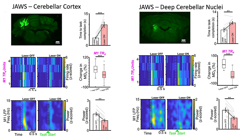
We have discovered that as animals master a skilled reaching task or a neuroprosthetic motor task, a distinctive pattern of slow, rhythmic electrical activity - low-frequency oscillations - emerges simultaneously in both brain areas, and only in animals that successfully learn the task. These coordinated rhythms appear to be the brain's way of synchronizing its motor circuits as a skill is refined. We have also found that the two regions within the cerebellum play distinct roles across learning: the outer layer of the cerebellum is critical during early, exploratory practice, while its deeper output structures are needed to maintain a well-learned skill. Together, these findings reveal the cerebellum as an active, essential partner in motor learning - not just a passive coordinator of movement.
Relevant papers: Abbasi et al., Science Advances, 2024; Fleischer et al., eNeuro, 2023, Ramanathan*, Guo*, Gulati* et al., Nature Medicine, 2018
Impaired modulation of motor cortex neuronal activity with cerebellar cortical and deep nuclear optogenetic stimulation.
Sleep: Where Memories Are Made (and Pruned)
We tend to think of learning as something that happens while we are practicing. But a large part of learning occurs while we sleep. During deep sleep, the brain quietly replays the experiences of the day, deciding which new patterns to strengthen into lasting memories and which to let fade - the process of memory consolidation.
Our lab has identified the precise sleep rhythms that govern this process for motor skills. We have shown that specific oscillations during non-REM (deep) sleep - particularly slow oscillations nested with sleep spindles - drive consolidation, while a separate set of rhythms (delta waves) actively promote forgetting. Using light-based tools that can switch individual neurons on or off with millisecond precision, we causally demonstrated that the sleeping brain is continuously making these keep-or-discard decisions - a process we call credit assignment. Strikingly, we have now extended this to the cerebellum: sleep spindles in both the motor cortex and the cerebellum recruit the same neurons that fired during daytime practice, and the coordination between these two regions during sleep predicts how much skill expertise is gained by the next morning. Sleep, in other words, is where the motor cortex and cerebellum cement their partnership.
Relevant papers: Fleischer*, Abbasi* and Gulati, eNeuro, 2024; Kim, Gulati and Ganguly, Cell, 2019; Gulati et al., Nature Neuroscience, 2017; Gulati et al., Nature Neuroscience, 2014
Two neurons recorded during two neuroprosthetic task - training sessions (left), and one cell’s optogenetic inhibition during intervening sleep (right).
Stroke Recovery as Re-Learning - and How to Accelerate It
A stroke - the sudden loss of blood flow to part of the brain - is in many ways a catastrophic interruption of the brain's learning machinery. The motor circuits that once coordinated skilled arm and hand movements are damaged, and recovery is fundamentally a process of re-learning: the surviving brain tissue must find new ways to coordinate movement, much like learning a skill for the first time, but starting from a disrupted foundation.
Our lab has found that the slow rhythmic oscillations that normally organize motor circuits during skilled movement are suppressed after stroke - and that recovery tracks their gradual restoration. Critically, we have shown that delivering precisely timed electrical stimulation during rehabilitation practice, locked to the moment these rhythms appear, significantly accelerates recovery of skilled movement in animal models of stroke. We have also shown that the cerebellum remains capable of driving neuroprosthetic even after stroke - its neurons can directly power a neuroprosthetic device as effectively as motor cortex neurons, even in animals with severe motor deficits. Our ongoing work is developing closed-loop cerebellar stimulation - delivered at the right moment during both waking practice and sleep - as a precision approach to rehabilitation that complements the growing interest in cerebellar deep brain stimulation for stroke.
Relevant papers: Rangwani et al., Cell Reports, 2025; Abbasi et al., Journal of Neural Engineering and Rehabilitation, 2021; Ramanathan*, Guo*, Gulati* et al., Nature Medicine, 2018
Cerebellar stimulation's effects on neural dynamics in stroke perilesional motor cortex.
From Brain to Muscle: Understanding Abnormal Movement After Stroke
Recovery from stroke is not just about relearning skilled movements. Many stroke survivors experience spasticity - involuntary muscle stiffness - and abnormal movement patterns in which muscles that should work independently become locked together in unwanted combinations. These patterns reflect disrupted coordination all the way from the cortex to the spinal cord.
Our lab studies this through the lens of muscle synergies: the coordinated patterns of muscle activation that the nervous system uses to simplify movement control. After stroke, these synergies become abnormal - and we suggest this may in part reflect changes in residual pathways directly damaged by the stroke, as well as increased influence of other pathways that now exert greater influence on spinal motor networks. Understanding how cortical pre-movement and movement states are shaped by the cortico-cerebellar circuits and how they compete with indirect pathways after a stroke and finally - how these relate to these spinal output patterns is an important open question that our lab is actively exploring. Muscle synergies also give us a practical, measurable outcome for evaluating whether our interventions are genuinely restoring normal motor coordination, not just improving gross performance scores.
Relevant papers: Abbasi and Gulati, Neuron, 2025; Godlove*, Gulati* et al., Annals of Clinical and Translational Neurology, 2016
Muscle synergies from both hind limbs showing that energy balances in a synergy are unilateral (represent muscles from one leg).
From Lab to Clinic: Finding Biomarkers to Guide Smarter Rehabilitation
Understanding the brain's recovery machinery in animal models is only meaningful if it translates to people. A key translational goal of our lab is to identify measurable signatures of recovery in the human brain after stroke - biomarkers that can tell us what state the brain is in, how well it is consolidating rehabilitation gains, and when it is most ready to benefit from stimulation.
Our clinical research uses bedside EEG - a non-invasive way to record the brain's electrical activity - to study motor training and sleep activity in hospitalized stroke patients. We have found that the healthy balance of sleep oscillations is profoundly disrupted after stroke: the restorative slow oscillation-spindle coupling that normally consolidates motor memories is suppressed on the stroke-affected side, while pathological delta-wave-nested patterns dominate. These asymmetries may help explain why stroke survivors often fail to consolidate the gains from their rehabilitation sessions. We are developing these sleep oscillation patterns as candidate biomarkers - measurable signals that can guide personalized, closed-loop stimulation therapies that enhance the brain's own recovery processes during both waking practice and sleep.
Relevant paper: Simpson*, Rangwani* et al., Frontiers in Neurology, 2023; Ramanathan*, Guo*, Gulati* et al., Nature Medicine, 2018; Godlove*, Gulati* et al., Annals of Clinical and Translational Neurology, 2016
Topographical map for detected NREM sleep oscillation density for stroke patients and their asymmetry based on stroke location.